
Cartilage Histology Quiz
How the Scales are Structured
Who Usually Takes This Test?
See How You Compare
Below is a preview of how scores are typically distributed across each scale.
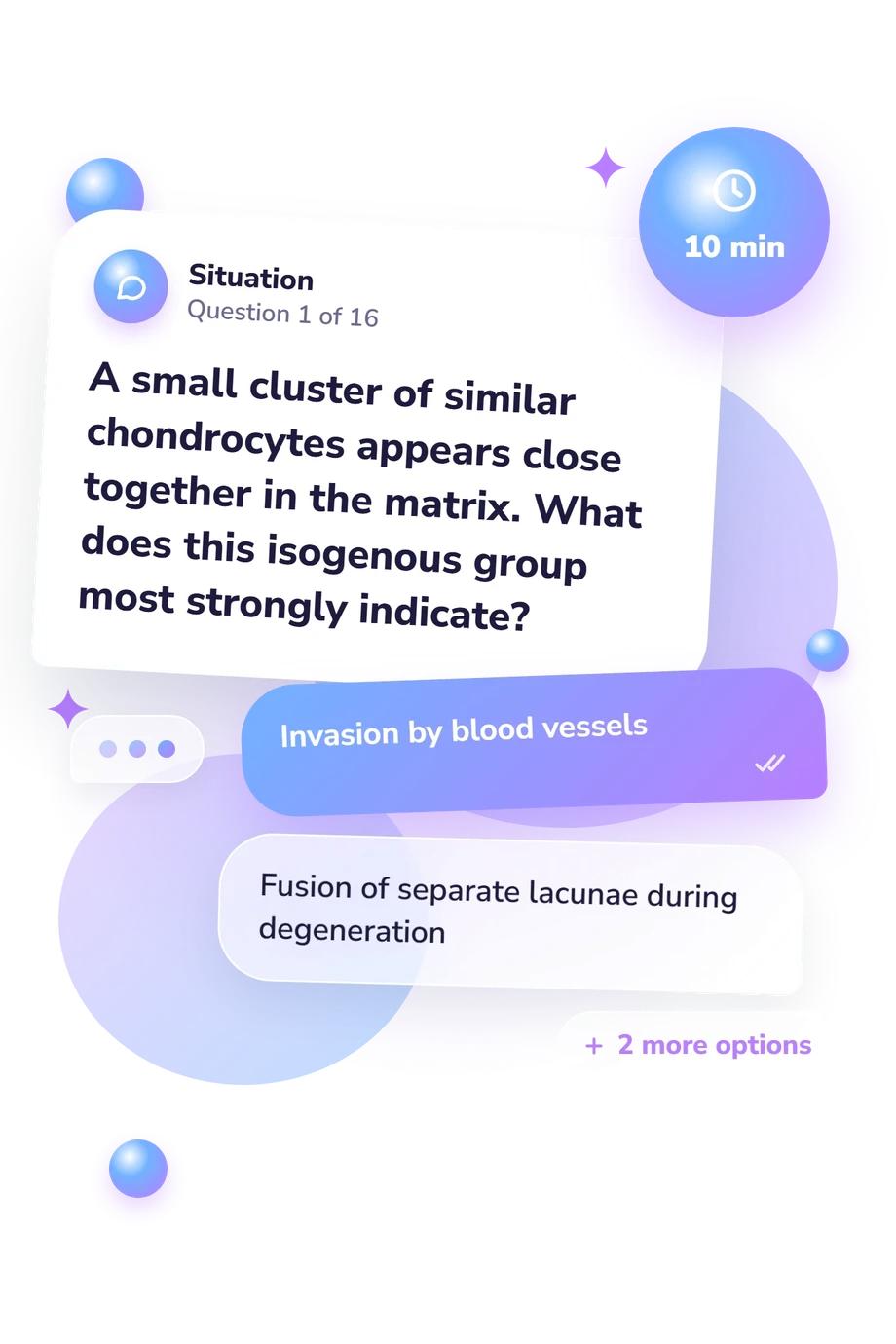
Example Questions From the Test
- Hyaline cartilage
- Fibrocartilage
- Dense irregular connective tissue
- Elastic cartilage
- Large deposits of neutral triglycerides
- Abundant calcium phosphate crystals
- Negatively charged sulfated glycosaminoglycans
- Concentrated elastic lamellae alone
- Chondrocytes cannot use nutrients carried in blood
- Cartilage is normally avascular
- The perichondrium blocks all exchange with surrounding tissue
- Lacunae are sealed by a mineralized wall
Cartilage Histology Quiz - Description and methodology of the test
Students and practitioners who want to consolidate microscopic anatomy can use this cartilage histology quiz, rather than a causes of world war 1 quiz, to connect tissue appearance with cell biology and function. Its result gives learners a gauge of knowledge and points toward broad areas for revision. This cartilage histology assessment keeps that review centred on cells, matrix, growth, repair, and tissue types, unlike a causes of ww1 quiz.
What This Assessment Explores
At its core, the assessment checks whether a learner can connect microscopic appearance with cartilage biology. It covers chondrocytes, lacunae, matrix, collagen and elastic fibers, vascularity, perichondrium, growth, repair, and major cartilage types. The focus suits students reviewing normal histology and readers testing applied recognition.
These cartilage histology questions test whether a feature is interpreted in context rather than memorized as an isolated label. The result is an overall knowledge level, not a world war 1 causes quiz score.
The result organizes performance through one scale:
- Cartilage Histology Knowledge (0-16) shows how accurately the respondent understands core cartilage features and applies them to histological scenarios. Foundational (0-6) signals partial recognition and a need to review cells, matrix, coverings, and tissue types; Developing (7-12) reflects useful working knowledge with distinctions or application still to strengthen; Strong (13-16) indicates accurate understanding of most core features.
Methodology and Scientific Basis
Each item uses a single-best-answer format: a brief histological or anatomical scenario is followed by four alternatives, and the respondent selects the option that best fits the structure or process. This tests recognition and application rather than personal experience.
Across the set, situations cover cell identification, matrix composition, growth, repair, staining, tissue classification, and functional location. The sequence links visible features with biological reasons, so careful reading matters as much as memorized terms.
As a cartilage histology test, it is conceptually consistent with Wheater’s Functional Histology, which supplies the core microscopic framework. Fox, Bedi, and Rodeo (2009) support the treatment of articular cartilage, including composition, avascular nutrition, and limited repair, while Benjamin and Ralphs (2004) add context for fibrocartilage cells and mechanical demands.
The scoring display reports performance through the displayed bands for this knowledge measure, not as a world war 1 quiz. Prior coursework, recall, image familiarity, and guessing can affect performance; the online format also cannot establish laboratory skill or clinical status. The result is best used to direct review toward uncertain distinctions and applied identification.
- O'Dowd G, Bell S, Wright S. Wheater's Functional Histology: A Text and Colour Atlas. 7th ed. Elsevier; 2024.
- Fox AJS, Bedi A, Rodeo SA. The Basic Science of Articular Cartilage: Structure, Composition, and Function. Sports Health. 2009;1(6):461-468. 10.1177/1941738109350438.
- Benjamin M, Ralphs JR. Biology of Fibrocartilage Cells. International Review of Cytology. 2004;233:1-45. 10.1016/S0074-7696(04)33001-9.
Updated: