
Female Reproductive System Histology Quiz
How the Scales are Structured
Who Usually Takes This Test?
See How You Compare
Below is a preview of how scores are typically distributed across each scale.
Example Questions From the Test
- Ovary
- Uterine cervix
- Vagina
- Uterine tube
- Primary follicle
- Primordial follicle
- Secondary antral follicle
- Corpus albicans
- Perimetrium
- Stratum basalis
- Myometrium
- Stratum functionalis
Female Reproductive System Histology Quiz - Description and methodology of the test
Students and readers who want to connect tissue appearance with organ function can use this assessment to check their grasp of microscopic anatomy. The female reproductive system histology quiz emphasizes recognition rather than isolated labels. Its questions move across organs and tissue changes. A reproductive histology quiz can support slide review before course exams. The result shows which structures and cycle-related patterns are secure and which need review.
What This Assessment Explores
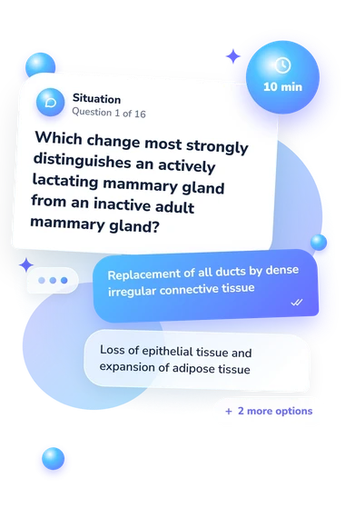
Rather than testing symptoms or personal health, this questionnaire asks whether learners can identify normal microscopic structures and connect them with location and function. It covers epithelial linings, ovarian follicles, endometrial phases, cervical and vaginal regions, placental villi, and mammary-gland activity. It suits students practising female reproductive histology.
The result is reported as Female Reproductive System Histology Knowledge (0-16). The scale measures recognition of reproductive tissues and their microscopic location-function relationships. Bands are Foundations to Revisit (0-6), Developing Recognition (7-12), and Strong Histology Knowledge (13-16).
The main themes are:
- Epithelial patterns show how lining cells identify organs.
- Ovarian structures show how follicles, atresia, corpus luteum, and medulla differ.
- Endometrial changes show how glands and stroma reflect cycle phases.
- Regional tissues show how cervix, vagina, placenta, and mammary gland are distinguished.
Methodology and Scientific Basis
Each item presents a histological description or comparison with four answer choices. The respondent selects the option matching the tissue, organ, cycle phase, or functional state. This tests applied recognition: epithelial type, folds, glands, stroma, vascularity, and follicular features must be considered together rather than as isolated clues.
Scores summarize performance across the questionnaire and direct review priorities. The result is identified as unvalidated and educational, so it offers study guidance rather than established norms or clinical cutoffs. A histology of the female reproductive system assessment can reveal knowledge gaps, but it cannot establish disease or assess a person’s body.
Conceptually, the content is consistent with Boyd et al. (2018), which describes microscopic organization of the female reproductive tract. Wheater, O'Dowd, Bell, Wright, Pawlina, and Ross provide accounts of normal structure and function. These sources support the subject matter, but the questionnaire is not presented as a standardized adaptation of a published instrument. Attention, teaching, answer selection, and context can affect performance, so difficult points should be checked against a histology text or course materials.
- Boyd, Kelli L., Atis Muehlenbachs, Mara H. Rendi, Rochelle L. Garcia, and Katherine N. Gibson-Corley. "Female Reproductive System." In Comparative Anatomy and Histology: A Mouse, Rat, and Human Atlas, 2nd ed., edited by Piper M. Treuting, Suzanne M. Dintzis, and Kathleen S. Montine, 303-334. Academic Press, 2018. 10.1016/B978-0-12-802900-8.00017-8
- O'Dowd, Geraldine, Sarah Bell, and Sylvia Wright. Wheater's Functional Histology: A Text and Colour Atlas. 7th ed. Elsevier, 2023.
- Pawlina, Wojciech, and Michael H. Ross. Histology: A Text and Atlas: With Correlated Cell and Molecular Biology. 8th ed. Wolters Kluwer, 2020.