
Tissue Quiz with Pictures
How the Scales are Structured
Who Usually Takes This Test?
See How You Compare
Below is a preview of how scores are typically distributed across each scale.
Example Questions From the Test
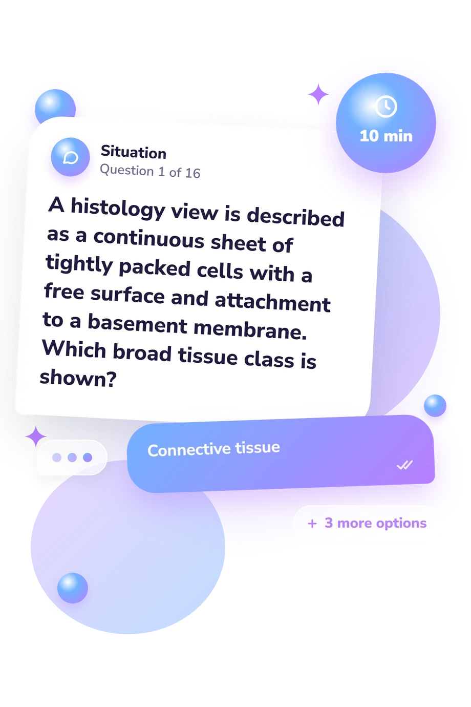
- Connective tissue
- Epithelial tissue
- Muscle tissue
- Nervous tissue
- Nervous tissue
- Blood
- Simple squamous epithelium
- Areolar connective tissue
- Smooth muscle
- Cardiac muscle
- Dense regular connective tissue
- Skeletal muscle
Tissue Quiz with Pictures - Description and methodology of the test
For learners revising microscopy, this assessment links visible structures with the names and functions of major tissue classes. Results from the tissue quiz with pictures show which recognition skills are secure and which deserve another look. An anatomy quiz on tissues with pictures suits students, instructors, and curious readers who want a focused way to connect histological clues with anatomy revision.
What This Assessment Explores
The questions examine how well a respondent links cell arrangement, matrix, surface specialization, and other structural details to tissue identity. Prompts use airways, skin, bladder, lung, tendon, blood, intestine, and skeletal muscle, so the tissue identification quiz tests whether knowledge transfers from a description or visual clue to specimen-based reasoning.
The content falls into four recurring groups:
- Epithelial tissue is identified through layers, cell shape, cilia, goblet cells, keratin, and stretch-related changes.
- Connective tissue is distinguished by matrix, fibers, cells, lacunae, and organization.
- Muscle tissue is separated by striations, branching, nuclei, and contraction pattern.
- Nervous tissue is recognized through large cell bodies, prominent nuclei, and branching processes.
The reported scale, Tissue Knowledge, runs from 0-16: Foundations to build (0-6), Developing recognition (7-12), and Strong tissue knowledge (13-16). It describes performance on tissue recognition, not intelligence or overall anatomy competence.
Methodology and Evidence Base
Each item presents a microscopic description or image-based clue with four answer choices. Correct responding requires combining cell shape, arrangement, extracellular matrix, nuclei, fibers, and specialized structures. The format rewards classification from observable features rather than recall of isolated terms.
Scoring summarizes tissue-recognition performance from selected answers. It can direct revision, but this online version has no stated norm group, reliability estimate, or diagnostic threshold. As a histology quiz with pictures, it is an educational snapshot shaped by study, memory, and familiarity with terminology.
Content is conceptually consistent with Pawlina and Ross (2020). Marieb and Smith (2024) and Betts et al. (2022) support anatomy and laboratory classification, while Heidger and colleagues (2002) is relevant to real and virtual imaging in teaching and testing. These works support the content and approach, not standardization of this questionnaire.
Interpret the result alongside actual errors: a missed item may reflect confusion between similar epithelia, difficulty reading structural clues, or a terminology gap. Study context, visual experience, and attention can affect performance, so the score is best used to choose the next review topic.
- Pawlina W, Ross MH. Histology: A Text and Atlas: With Correlated Cell and Molecular Biology. 8th ed. Philadelphia, PA: Wolters Kluwer; 2020.
- Marieb EN, Smith LA. Human Anatomy & Physiology Laboratory Manual, Main Version, Global Edition. 12th ed. Pearson; 2024.
- Betts JG, Young KA, Wise JA, et al. Anatomy and Physiology 2e. Houston, TX: OpenStax; 2022.
- Heidger PM Jr, Dee F, Consoer D, Leaven T, Duncan J, Kreiter C. Integrated approach to teaching and testing in histology with real and virtual imaging. The Anatomical Record. 2002;269(2):107-112. 10.1002/ar.10078
Updated: