
Brachial Plexus Quiz
How the Scales are Structured
Who Usually Takes This Test?
See How You Compare
Below is a preview of how scores are typically distributed across each scale.
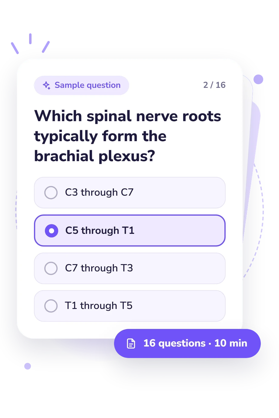
Example Questions From the Test
- C3 through C7
- C5 through T1
- C7 through T3
- T1 through T5
- Ulnar nerve
- Musculocutaneous nerve
- Radial nerve
- Axillary nerve
- Musculocutaneous nerve
- Median nerve
- Axillary nerve
- Ulnar nerve
Brachial Plexus Quiz - Description and methodology of the test
Designed for learners reviewing upper-limb anatomy, this assessment helps connect classroom knowledge with the nerve deficits that follow common lesions. The brachial plexus quiz is useful for students who want to check whether recent revision is secure and decide which topics deserve another pass. Its result can turn uncertainty into a focused brachial plexus study plan, especially when a broad anatomy review needs a clear starting point.
What This Assessment Measures
The questions examine whether you can map the plexus from roots through trunks, divisions, cords, and terminal branches, then apply that map to injury patterns. They also test relationships around the axillary artery, sensory territories, motor actions, and root contributions to named nerves. Together, these structure-identification and lesion-localization questions make a brachial plexus anatomy review practical: you connect a lesion pattern with the most likely affected nerve or segment.
The result is educational: it summarizes how consistently your answers matched the tested anatomy and guides review. The Brachial Plexus Knowledge scale runs from 0 to 16 and has three result bands:
- Foundations to Review (0-6) indicates that the pathway, branches, or relationships need strengthening.
- Developing Knowledge (7-12) shows a workable grasp; reviewing branch origins and lesion localization remains useful.
- Strong Knowledge (13-16) reflects a command of organization and clinical applications.
Methodology and Evidence Base
Each item presents an anatomical or clinical prompt with four answer choices. One point is assigned for each correct answer, so the total is based on keyed responses. Across the set, structure-identification and clinical-localization items link Erb or Klumpke patterns, winged scapula, sensory changes, and extension weakness to a root, trunk, cord, or nerve.
The anatomical relationships tested here are consistent with the anatomy described by Neto et al. (2022). Leijnse et al. (2020) helps explain developmental variation in plexus organization, while Sakellariou et al. (2014) supplies clinical context for adult injury examination and localization. These publications provide anatomical and clinical context for the tested relationships; they are not instruments used by this questionnaire.
As a brachial plexus anatomy test, it is best interpreted as a focused self-check. Recall, wording, guessing, and current study context can affect performance. The bands describe educational knowledge only; they do not diagnose an injury or establish clinical competence. Use missed relationships to choose what to review next, checking them against a reliable anatomy source as part of further study.
- Neto JHS, Neto BC, Eiras ABD, Botelho RHS, de Carmo JMM, Passos MARF. The 2-Dimensional and 3-Dimensional Anatomy of the Adult Brachial Plexus Divisions and Cords. Hand (N Y). 2022;17(1):50-54. 10.1177/1558944720906510
- Leijnse JN, de Bakker BS, D'Herde K. The brachial plexus - explaining its morphology and variability by a generic developmental model. Journal of Anatomy. 2020;236(5):862-882. 10.1111/joa.13123
- Sakellariou VI, Badilas NK, Mazis GA, Stavropoulos NA, Kotoulas HK, Kyriakopoulos S, Tagkalegkas I, Sofianos IP. Brachial Plexus Injuries in Adults: Evaluation and Diagnostic Approach. ISRN Orthopedics. 2014;2014:726103. 10.1155/2014/726103
Updated: