
Dental Radiography Quiz
How the Scales are Structured
Who Usually Takes This Test?
See How You Compare
Below is a preview of how scores are typically distributed across each scale.
Example Questions From the Test
- Failure to place the embossed identification dot occlusally
- Excessive horizontal angulation
- Insufficient receptor coverage
- Movement of the patient, receptor, or tubehead during exposure
- Bitewing
- Periapical
- Panoramic
- Posterior occlusal
- Increasing image-receptor sensitivity
- Adding display contrast after exposure
- Using rectangular instead of circular collimation
- Using a longer exposure time
Dental Radiography Quiz - Description and methodology of the test
Students, trainees, and practitioners revisiting core imaging decisions can use the dental radiography quiz to decide whether their foundations are ready for further study. As a dental radiography assessment, it asks respondents to interpret radiographic appearances and choose the most likely explanation or appropriate principle. The result offers an educational snapshot for judging readiness to continue reviewing.
What This Assessment Explores
Those checking core imaging knowledge can use this dental radiography practice test to connect visible radiographic features with likely technical causes and appropriate projections. It suits learners, trainees, and practitioners reviewing dental imaging principles.
The displayed scale is Dental Radiography Knowledge (0-16): Foundations to Review (0-6), Developing Knowledge (7-12), and Strong Knowledge (13-16). These bands give a broad indication of command of the sampled material, from foundations needing review to stronger knowledge.
The questions address four recurring content areas:
- Positioning and geometry show how receptor placement and beam alignment affect coverage.
- Image quality links blur, cone cuts, missing anatomy, and overlap with likely causes.
- Projections and anatomy identify what periapical, bitewing, and panoramic views display.
- Protection and quality assurance address justification, operator safety, exposure, and repeats.
One overall result represents these themes.
Methodology and Scientific Basis
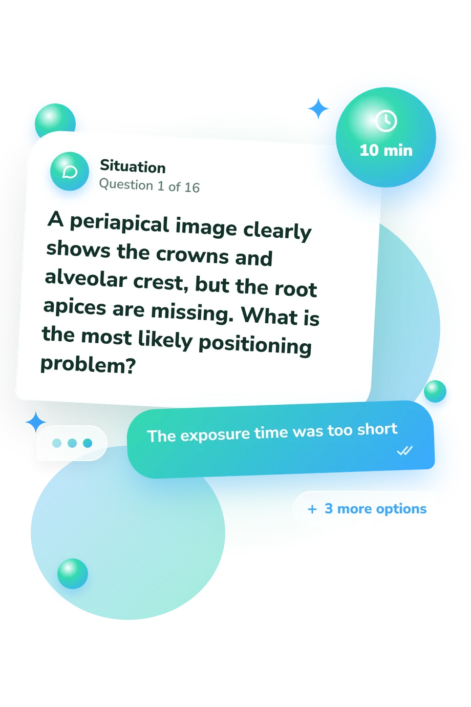
Each item presents a situation with four answer choices. Respondents select the option that best explains a positioning error, image appearance, projection, geometric relationship, protection practice, or quality-assurance decision. These radiographic interpretation questions emphasize applied recognition rather than free-form recall.
One displayed scale is reported, and no separate scores are shown for geometry, projections, image quality, or protection.
The framework is conceptually consistent with Iannucci and Howerton (2021) on technique, image formation, projections, and radiation protection. Patient selection and justification correspond with Benavides et al. (2026); repeat reduction and image quality align with Goren et al. (2000). This is a separate educational questionnaire, not an adaptation of those reference works, recommendations, or the published quality-assurance study.
An overall result can guide revision by indicating broad knowledge across the sampled material, not a particular weak topic. Because the score is unvalidated and reflects sampled educational items, it may vary with study, attention, wording, and self-awareness; the highest band does not establish clinical competence, and exposure and safety decisions still follow local procedures, manufacturer guidance, and qualified supervision.
- Iannucci, Joen M., and Laura Jansen Howerton. Dental Radiography: Principles and Techniques. 6th ed. Elsevier, 2021. ISBN 978-0-323-69550-3.
- Benavides, Erika, Joseph R. Krecioch, Trishul Allareddy, et al. "American Dental Association and American Academy of Oral and Maxillofacial Radiology patient selection for dental radiography and cone-beam computed tomography: Clinical recommendations." Journal of the American Dental Association 157, no. 1 (2026): 20-35.e5. 10.1016/j.adaj.2025.10.013.
- Goren, A. D., R. C. Lundeen, S. T. Deahl II, et al. "Updated quality assurance self-assessment exercise in intraoral and panoramic radiography." Oral Surgery, Oral Medicine, Oral Pathology, Oral Radiology, and Endodontics 89, no. 3 (2000): 369-374. 10.1016/S1079-2104(00)70104-1.
Updated: