
Epidermis Layers Quiz
How the Scales are Structured
Who Usually Takes This Test?
See How You Compare
Below is a preview of how scores are typically distributed across each scale.
Example Questions From the Test
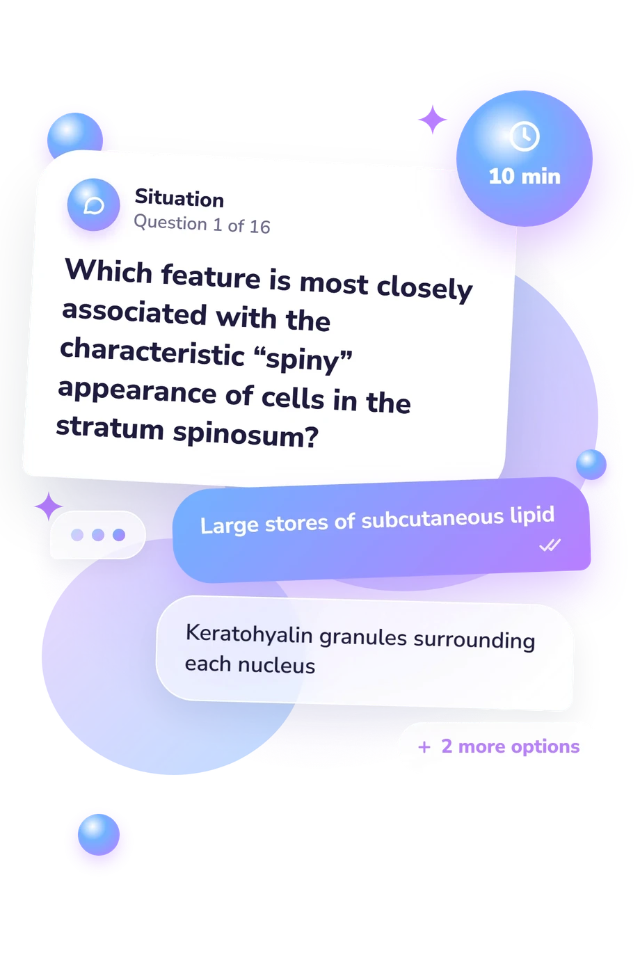
- Large stores of subcutaneous lipid
- Keratohyalin granules surrounding each nucleus
- Desmosomal junctions between keratinocytes
- A continuous sheet of dermal capillaries
- Basale → granulosum → spinosum → corneum
- Basale → spinosum → granulosum → corneum
- Spinosum → basale → granulosum → corneum
- Corneum → granulosum → spinosum → basale
- Prominent keratohyalin granules
- Dense networks of blood vessels
- Clusters of actively dividing stem cells
- Bundles of melanin-producing cilia
Epidermis Layers Quiz - Description and methodology of the test
Students, trainees, and readers who want to check their grasp of skin histology can use this assessment to connect structure with function. Rather than surveying dermatology, the epidermis layers quiz concentrates on the strata that form the skin’s epithelial barrier. It is useful for anatomy review, microscopy preparation, and checking whether memorized labels support reasoning. The result of this quiz identifies concepts about the epidermis worth reviewing.
What This Assessment Explores
As a quiz about the layers of the epidermis, this test measures epidermal strata knowledge in tissue context. Its questions ask you to sequence those layers and connect their features with skin function, including defining cells and structures, thin-versus-thick skin, and roles in renewal, pigmentation, barrier formation, and superficial injury. It suits histology and anatomy review.
The displayed scale is Epidermis Layers Knowledge (0-16). Each correct answer earns one point, with Foundations to build (0-6), Developing knowledge (7-12), and Strong knowledge (13-16). The bands describe command of the tested content, not overall biological ability.
The recurring learning tasks are:
- Layer order shows whether you can trace keratinocytes from base to surface.
- Microscopic features show whether you can match granules, junctions, and cell shapes to layers.
- Regional and functional comparisons show whether you can distinguish thick from thin skin and link strata to renewal, pigmentation, barriers, and nutrient supply.
Methodology and Scientific Basis
Each item uses single-best-answer multiple choice with four alternatives. The scenarios ask you to identify a layer, order strata, recognize a microscopic feature, or infer a consequence of normal epidermal structure. This checks recall alongside simple application rather than personal opinions.
In practical terms, this epidermal strata quiz uses direct answer-key scoring: a correct choice contributes one point, and the total summarizes performance across the content domains. Its content is conceptually consistent with Simpson et al. (2011), whose work emphasizes keratinocyte differentiation, adhesion, outward migration, and epidermal homeostasis. Pawlina and Ross, together with Mescher, supply standard histological descriptions of the strata, their cells, and thick-versus-thin skin differences. Mascré et al. (2012) provide primary research on progenitor contributions to epidermal maintenance, a context relevant to the renewal items but not a validation study of this questionnaire.
The bands are content-based result categories, so the result is best read as a study signal. Performance can vary with recent revision or interpretation of a microscopy-style prompt; practical slide work or course assessment can add context when judging mastery.
- Simpson CL, Patel DM, Green KJ. Deconstructing the skin: cytoarchitectural determinants of epidermal morphogenesis. Nature Reviews Molecular Cell Biology. 2011;12(9):565-580. 10.1038/nrm3175
- Pawlina W, Ross MH. Histology: A Text and Atlas: With Correlated Cell and Molecular Biology. 8th ed. Philadelphia: Wolters Kluwer Health; 2020.
- Mescher AL. Junqueira's Basic Histology: Text and Atlas. 16th ed. New York: McGraw-Hill Education; 2021.
Updated: