
Muscle Fiber Quiz
How the Scales are Structured
Who Usually Takes This Test?
See How You Compare
Below is a preview of how scores are typically distributed across each scale.
Example Questions From the Test
- Fascicles
- Sarcomeres
- Terminal cisternae
- Motor units
- A myosin head
- A troponin molecule
- A calcium ion
- A tropomyosin strand
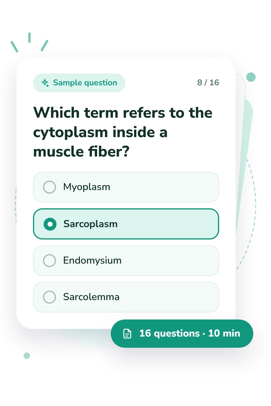
- Myoplasm
- Sarcoplasm
- Endomysium
- Sarcolemma
Muscle Fiber Quiz - Description and methodology of the test
Students reviewing anatomy, instructors checking foundational understanding, and curious learners can use this skeletal muscle anatomy quiz to identify how well they recognize the organization of skeletal muscle fibers. The muscle fiber quiz indicates whether your command of tested anatomy is foundational, developing, or strong. It is especially useful before studying excitation-contraction coupling or interpreting labeled diagrams.
What This Assessment Measures
Questions test whether learners connect names, locations, and functions across skeletal muscle structure. Several prompts give learners practice identifying labeled muscle-fiber regions, so this muscle fiber anatomy quiz reinforces distinctions among terms that are easy to confuse. The thematic areas are:
- Myofilament organization shows whether sarcomere landmarks are related correctly.
- Membrane and calcium systems identify routes and stores used in excitation-contraction coupling.
- Contractile proteins and shortening reveal understanding of cross-bridge action and band changes.
- Fiber hierarchy and coverings show whether structures are ordered from components to whole muscle.
The result uses one displayed dimension: Muscle Fiber Knowledge, scored from 0-16 correct. Foundation to Build covers 0-6, Developing Command covers 7-12, and Strong Command covers 13-16. These bands describe foundational, developing, or strong command of the tested anatomy. The assessment suits learners preparing for physiology or health-related coursework; it does not measure strength or clinical muscle function.
Methodology and Evidence Base
Each item in this muscle fiber anatomy test presents a question with four answer choices, asking the learner to select the term or description that best fits the anatomical situation. The format tests recognition and discrimination rather than free-response explanation; distractors may come from neighboring structures, so careful comparison matters. Several items cover Z discs, A bands, I bands, H zones, M lines, and filament overlap, requiring discrimination among closely related structures.
Scoring counts correct answers and reports a study snapshot. Betts et al. (2022) supplies the core anatomy framework for fibers, sarcomeres, membranes, connective tissues, and excitation-contraction structures. Squire (2016) explains sliding-filament contraction and sarcomere dynamics, while Flucher, Weiss, and Grabner (2002) supports triads in excitation-contraction coupling. The online questionnaire is conceptually consistent with the cited anatomy and contraction literature, and its displayed bands are learning descriptors rather than population norms.
Use the result to plan review: missed items point to anatomy terms or relationships that need another look, while a high result indicates strong performance across the tested material.
- J. Gordon Betts, Kelly A. Young, James A. Wise, Eddie Johnson, Brandon Poe, Dean H. Kruse, Oksana Korol, Jody E. Johnson, Mark Womble, and Peter DeSaix. Anatomy and Physiology 2e. Houston, TX: OpenStax, 2022, Section 10.2, “Skeletal Muscle.”
- John M. Squire. “Muscle contraction: Sliding filament history, sarcomere dynamics and the two Huxleys.” Global Cardiology Science & Practice 2016, no. 2 (2016): e201611. 10.21542/gcsp.2016.11
- Bernhard E. Flucher, Regina G. Weiss, and Manfred Grabner. “Cooperation of two-domain Ca2+ channel fragments in triad targeting and restoration of excitation-contraction coupling in skeletal muscle.” Proceedings of the National Academy of Sciences 99, no. 15 (2002): 10167-10172. 10.1073/pnas.122345799
Updated: