
Eye Anatomy Quiz
How the Scales are Structured
Who Usually Takes This Test?
See How You Compare
Below is a preview of how scores are typically distributed across each scale.
Example Questions From the Test
- Canaliculi → lacrimal sac → nasolacrimal duct
- Nasolacrimal duct → canaliculi → lacrimal sac
- Canaliculi → nasolacrimal duct → lacrimal sac
- Lacrimal sac → canaliculi → nasolacrimal duct
- Choroid → sclera → retina
- Retina → choroid → sclera
- Sclera → choroid → retina
- Sclera → retina → choroid
- Orbicularis oculi muscle
- Superior rectus muscle
- Iris dilator muscle
- Ciliary muscle
Eye Anatomy Quiz - Description and methodology of the test
Students, trainees, educators, and curious learners can use this eye quiz to check their understanding of ocular structures and pathways. An overall anatomy knowledge result gives learners a practical way to gauge their command of core material and organize further study. For learners preparing to revise or consolidate anatomy, the eye anatomy quiz offers a focused measure across essential eye structures and functions.
What This Assessment Explores
Questions cover the eye's outer protection, tear drainage, internal chambers and coats, focusing structures, pupil control, retinal organization, photoreceptors, visual fields, and the optic nerve. It is a quiz about the parts of the eye that connects names with locations, functions, and sequences.
The content areas are:
- Surface and drainage structures show how protection and tear movement support the eye.
- Chambers, coats, and focusing structures show how the globe is organized and light is controlled.
- Retinal and optic pathways show how light becomes neural information and reaches the brain.
The catalogue reports one result scale: Eye Anatomy Knowledge, 0-16. Foundation to build (0-6) indicates that basic recognition is present but review would help; Developing knowledge (7-12) reflects understanding of many core concepts with some gaps; Strong knowledge (13-16) reflects a well-developed command of major structures and relationships.
Methodology and Scientific Basis
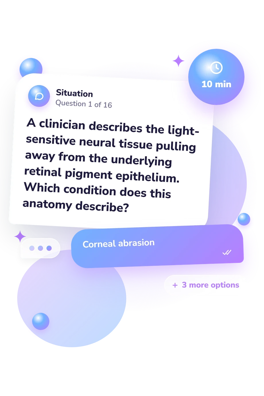
The questionnaire combines direct anatomy questions with short clinical-style descriptions and ordering tasks, followed by four answer choices. The items test naming, location, ordering, and functional relationships through recognition and application rather than written recall.
This questionnaire is conceptually consistent with the educational focus of Soylu and colleagues' ophthalmic anatomy learning study. Ansari and Nadeem’s atlas and Moore, Dalley, and Agur’s anatomy text provide reference frameworks for structures, pathways, and terminology. These sources support the subject matter, not the online questionnaire's psychometric properties.
Scoring counts correct responses, with one point for each correct answer, and the result is read through the three catalogue bands stated above. Because this anatomy of the eye quiz focuses on core structures and relationships, the total score can guide revision toward the topics represented by missed items. No published norms or validation data have been established for this online questionnaire, so performance can reflect guessing, recent teaching, memory, and familiarity with anatomical language.
- Soylu, Ayşe; Üçgül, Ahmet Yücel; Alkan, Raşid; Aktepe-Teke, Gülnaz; Akdağ, İhsan; Özkan, Ecem; Atalay, Hatice Tuba; Uysal, Betül Seher; Tarlan, Berçin; Kıyak, Yavuz Selim; Bilgihan, Kamil; Peker, Tuncay Veysel. "Virtual reality enhances ophthalmic anatomy learning: experimental evidence on comparable outcomes with peer and expert facilitators." BMC Medical Education, 26, 1409 (2026). 10.1186/s12909-026-09659-w
- Ansari, Mohammad Wakeel, and Ahmed Nadeem. Atlas of Ocular Anatomy. Springer, 2016. 10.1007/978-3-319-42781-2
- Moore, Keith L., Arthur F. Dalley II, and Anne M. R. Agur. Moore's Clinically Oriented Anatomy. 9th ed. Wolters Kluwer, 2023.
Updated: